







Chapter 4IMAT Biology
The Plasma Membrane
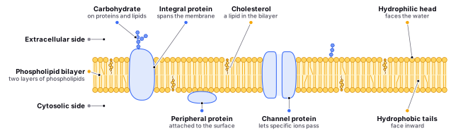
Imagine a city surrounded by a protective wall that separates the bustling activity inside from the outside world. In the same way, the plasma membrane is a barrier that encloses the busy and complex inner workings of the cell from the surrounding environment. The plasma membrane, also known as the cell membrane, is a thin and flexible barrier that surrounds the cell and separates the interior of the cell from the external environment. It is made up of a phospholipid bilayer, which is a double layer of phospholipid molecules. The phospholipid bilayer is composed of two layers of phospholipid molecules arranged with their hydrophobic tails facing inward and their hydrophilic heads facing outward.
But the plasma membrane is not just a static wall - it is a dynamic structure constantly in motion, like a dance floor with different types of dancers moving and changing positions. This is where the fluid mosaic model comes in, describing the plasma membrane as a fluid-like mosaic of different types of molecules, like a painting made up of colorful tiles. The fluid mosaic model is a model that suggests that the plasma membrane is a dynamic structure that is composed of a mosaic of different types of molecules, including phospholipids, proteins, and carbohydrates. These molecules are constantly moving and rearranging themselves within the membrane, giving it a fluid-like quality.
And just like how the city wall has gates for people and goods to enter and exit, the plasma membrane has proteins that act as gatekeepers, allowing molecules to pass through and communicating with other cells. These proteins can be found either embedded within the membrane or attached to its surface, like players on a sports team working together for the benefit of the whole. The fluid mosaic model also proposes that proteins in the plasma membrane play important roles in the transport of molecules across the membrane, as well as in cell-cell communication and signaling. The proteins can be either integral proteins, which are embedded within the phospholipid bilayer, or peripheral proteins, which are attached to the surface of the membrane.
Find it in the figure
Coming soon for figure 4.1
Section 4.1
The Concentration Gradient
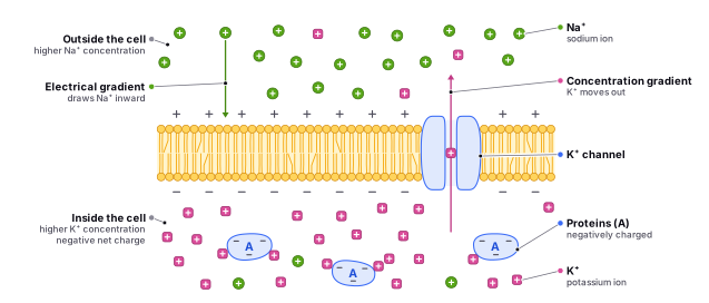
Concentration gradient is a term used to describe the difference in the concentration of a substance between two regions. In the context of cell biology, it refers to the difference in the concentration of a substance on either side of the cell membrane. This difference in concentration is what drives the movement of substances across the membrane.
Before delving into the various channels involved in the plasma membrane, it is important to have a basic understanding of concentration gradients. The concept of concentration gradient is fundamental to understanding how substances move across the membrane and the role of different channels in this process.
Substances move across the membrane from areas of high concentration to areas of low concentration, in order to reach a state of equilibrium. This movement is driven by the concentration gradient, which acts like a force that pushes the substance from an area of high concentration to an area of low concentration.
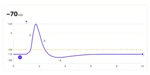
The concentration gradient is an important concept to be aware of because it is a driving force for many physiological processes. For example, the movement of ions across the membrane is essential for the generation and propagation of action potentials in neurons and muscle cells. The concentration gradient of ions across the membrane plays a critical role in establishing the resting potential of cells, which is necessary for the transmission of electrical signals.
Additionally, understanding concentration gradients is important for understanding the role of different channels in the plasma membrane. Channels are specialized proteins that allow the movement of substances across the membrane. The specific types of substances that can pass through these channels are determined by the properties of the channels themselves, as well as the concentration gradient of the substance across the membrane.
For example, ion channels allow the movement of ions across the membrane in response to changes in the concentration gradient of the ion. Gated channels open or close in response to specific stimuli, such as changes in voltage or the binding of a specific molecule. Pore-forming channels, on the other hand, are formed by the oligomerization of multiple channel proteins and can be either selective or non-selective in terms of the types of substances that can pass through.
In summary, understanding the concept of concentration gradient is essential for understanding how substances move across the plasma membrane and the role of different channels in this process. It is an important topic to be aware of before delving into the details of the different types of channels involved in the plasma membrane.
Find it in the figure
Coming soon for figure 4.2
Section 4.2
Membrane Proteins
Integral proteins and peripheral proteins are two types of proteins found in the plasma membrane.
Integral Proteins
Integral proteins are embedded within the phospholipid bilayer of the plasma membrane and are an integral part of the membrane structure. They are also known as trans-membrane proteins because they span the entire thickness of the membrane, with one end inside the cell and the other end outside the cell. Integral proteins have hydrophobic regions that interact with the hydrophobic tails of the phospholipid molecules in the membrane, allowing them to be integrated into the membrane structure.
There are several types of integral proteins, including:
Channel proteins: These proteins create channels through the membrane that allow specific ions or small molecules to pass through.
Channel Proteins
Channel proteins are integral membrane proteins that allow ions and small molecules to pass through the membrane by creating a channel through which the substances can pass. These proteins are important in the movement of substances across the membrane, which is essential for maintaining homeostasis within cells and tissues.
There are several types of channel proteins, including:
Ion Channels
Ion channels: These proteins allow ions to pass through the membrane. Ion channels can be either selective or non-selective, meaning that they can allow specific ions to pass through or any ion to pass through, respectively.
Ion channels are a type of channel protein that allow ions to pass through the membrane. Ion channels are integral membrane proteins, meaning that they are embedded within the lipid bilayer of the cell membrane. They are composed of proteins called subunits, which form a channel through which ions can pass.
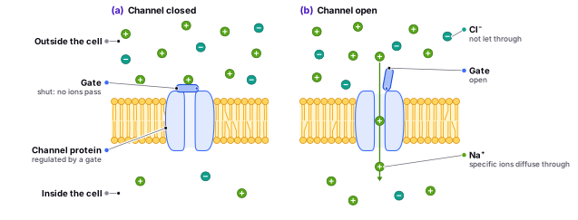
Ion channels can be either selective or non-selective. Selective ion channels allow specific ions to pass through the channel, while non-selective ion channels allow any ion to pass through. Selective ion channels are specific to certain ions, such as sodium, potassium, calcium, or chloride ions.
Ion channels can also be classified based on their activation properties. Some ion channels are always open, while others are gated, meaning that they can open or close in response to specific stimuli. For example, some ion channels are activated by changes in the voltage across the membrane, while others are activated by the binding of a specific molecule.
One example of an ion channel is the potassium leak channel, which is involved in maintaining the resting membrane potential of nerve cells. Another example is the calcium channel, which is involved in muscle contraction and neurotransmitter release. Chloride channels are found in the kidneys and are involved in the regulation of salt and water balance.
Ion channels are found in many different locations within the body, including nerve cells, muscle cells, and epithelial cells. In nerve cells, ion channels play a crucial role in the generation and transmission of action potentials. In muscle cells, ion channels are involved in muscle contraction. In epithelial cells, ion channels are involved in the transport of ions and water across cell membranes.
Find it in the figure
Coming soon for figure 4.3
Gated Channels
Gated channels are important proteins that control the flow of ions across the cell membrane in response to specific stimuli. They act like tiny gates, opening and closing to allow ions to pass through in a regulated manner.
One example of gated channels is voltage-gated channels. These channels are activated by changes in the voltage across the membrane, and are important in the generation and propagation of action potentials in neurons and muscle cells. An analogy to explain how voltage-gated channels work is to imagine a gate that opens when a certain amount of pressure is applied to it. In the case of voltage-gated channels, the gate opens when a certain voltage threshold is reached.
Another type of gated channel is ligand-gated channels. These channels are activated by the binding of a specific molecule, called a ligand, to the channel protein. Ligand-gated channels are important in the transmission of signals between cells and in the regulation of enzyme activity. An analogy to explain how ligand-gated channels work is to imagine a gate that opens when a specific key is inserted into the lock. In the case of ligand-gated channels, the key is the ligand that binds to the channel protein, causing the gate to open.
Mechanically-gated channels are another type of gated channel that are activated by physical stimuli, such as pressure or stretch. Mechanically-gated channels are important in the regulation of blood pressure and in the detection of sensory stimuli. An analogy to explain how mechanically-gated channels work is to imagine a gate that opens when a certain amount of force is applied to it. In the case of mechanically-gated channels, the gate opens when the membrane is stretched or compressed by physical stimuli.
Gated channels play a crucial role in many physiological processes, including the transmission of signals within and between cells, the regulation of enzyme activity, and the generation and propagation of action potentials. Without gated channels, the proper functioning of many biological processes would be disrupted.
Leak Channels
Leak channels are present in many types of cells, including neurons, muscle cells, and epithelial cells. In neurons, leak channels help to maintain the resting potential of the cell, which is essential for the transmission of action potentials. The resting potential of a neuron is typically around -70 millivolts, which is established by the concentration gradients of ions across the cell membrane. Leak channels contribute to the passive movement of ions down their concentration gradients, which helps to maintain the resting potential. The pore of the channel is lined with amino acids that interact with ions as they pass through the channel. The selectivity of the channel for specific ions is determined by the size and charge of these amino acids. While leak channels are non-selective, they do exhibit some preference for certain ions based on their size and charge.
Leak channels also play a role in regulating cell volume. For example, in kidney cells, leak channels help to regulate the osmotic pressure inside the cell by allowing the passive movement of ions across the membrane. This helps to prevent the cell from swelling or shrinking in response to changes in the extracellular environment.
An analogy for leak channels is a leaky faucet. Just as a leaky faucet allows water to pass through continuously, leak channels allow ions to pass through the membrane continuously. However, unlike a leaky faucet, leak channels are essential for maintaining the normal functioning of cells and tissues.
In summary, leak channels are always open and allow ions to pass through the membrane continuously, which helps to maintain the resting potential of cells and regulate cell volume. While leak channels may seem insignificant compared to gated channels, they play an important role in many physiological processes and are essential for the normal functioning of cells and tissues.
Pore-forming Channels
Pore-forming channels are a type of channel protein that are formed by the oligomerization, or the grouping together, of multiple channel proteins. Pore-forming channels can be either selective or non-selective, depending on the specific proteins that form the channel. These channels are characterized by a pore or channel that is formed by the oligomerization of the protein subunits. The channel is typically lined with hydrophilic residues, which allows it to interact with and transport ions or other small molecules.
Pore-forming channels play an essential role in many physiological processes, including the transport of ions and small molecules across the membrane, the regulation of cell volume, and the maintenance of ion gradients across the membrane. Some examples of pore-forming channels include:
- Voltage-gated potassium channels: These channels are activated by changes in the voltage across the membrane and allow the movement of potassium ions through the membrane. Voltage-gated potassium channels are important in the regulation of the resting potential of cells and in the repolarization phase of action potentials.
- Connexons: These channels are formed by the oligomerization of six connexin proteins and allow the movement of ions and small molecules between cells. Connexons are important in the communication between cells and in the regulation of enzyme activity.
- Aquaporins: These channels are selective for the movement of water molecules and allow the rapid movement of water across the membrane. Aquaporins are important in the regulation of water balance in the body.
- Porins: These channels are formed by the oligomerization of multiple porin proteins and allow the movement of small molecules through the outer membrane of bacteria. Porins are important in the uptake of nutrients and the elimination of waste products in bacteria.
An analogy for pore-forming channels could be a pipe or a tunnel that allows the movement of water or other substances. Just as a pipe allows the movement of water from one location to another, pore-forming channels allow the movement of ions or other small molecules across the membrane.
In summary, pore-forming channels are a type of channel protein that form a pore or channel through which ions or small molecules can move across the membrane. These channels play an essential role in many physiological processes, including the regulation of the resting potential of cells, the communication between cells, and the maintenance of ion gradients across the membrane.
Aquaporins
Aquaporins are highly selective channels that only allow the passage of water molecules through the membrane, preventing the passage of other small molecules and ions. They are able to selectively filter water due to their specific structural features. The narrowest part of the channel, known as the selectivity filter, is formed by a chain of amino acids that are arranged in a specific manner to create a pore that allows only water molecules to pass through. The presence of hydrophobic residues lining the pore also helps to prevent the passage of ions and other small molecules.
Aquaporins are important in many physiological processes. For example, they play a crucial role in the kidney's ability to reabsorb water from the urine and return it to the body. In the absence of aquaporins, the urine produced by the kidneys would be very dilute and the body would lose water rapidly, leading to dehydration.
An analogy for aquaporins can be a water slide at a water park. Just as a water slide allows only water to pass through while preventing the passage of other objects, aquaporins allow only water to pass through their channel while excluding other molecules and ions.
There are several types of aquaporins, including:
Aquaporin 0 (AQP0): AQP0 is found in the lens of the eye and is important in maintaining the transparency of the lens.
Aquaporin 1 (AQP1): AQP1 is found in the endothelial cells that line the blood vessels and is important in the regulation of blood pressure.
Aquaporin 2 (AQP2): AQP2 is found in the renal tubules of the kidney and is important in the regulation of water balance in the body.
Aquaporin 4 (AQP4): AQP4 is found in the brain and is important in the regulation of brain edema.
Aquaporins are important in maintaining proper hydration and electrolyte balance within cells. Dysfunction of aquaporins can lead to various diseases and disorders, including diabetes insipidus, a condition characterized by excessive thirst and the excretion of large amounts of dilute urine. Therefore, understanding the structure and function of aquaporins is important for the development of treatments for these conditions.
In addition to these types of channel proteins, there are also several subtypes of channel proteins that can be classified based on their specific functions. For example, there are channels that specifically allow the movement of calcium ions, potassium ions, or sodium ions.
Carrier Proteins
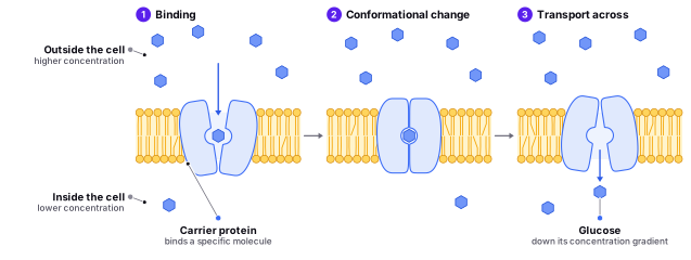
Carrier proteins are integral membrane proteins that bind to specific molecules and transport them across the membrane. These proteins undergo conformational changes upon binding of the specific molecule, which allows the molecule to be transported across the membrane.
Uniporters are carrier proteins that transport a single type of substance across the membrane. Uniporters can be either passive or active. Passive uniporters transport substances down their concentration gradient, while active uniporters require energy to transport substances against their concentration gradient. One example of a uniporter is the , which transports molecules across the plasma membrane.
Symporters are carrier proteins that transport two types of substances across the membrane in the same direction. Symporters can be either passive or active. Passive symporters transport substances down their concentration gradient, while active symporters require energy to transport substances against their concentration gradient. One example of a symporter is the sodium-glucose cotransporter, which transports both sodium ions and glucose molecules across the plasma membrane.
Antiporters are carrier proteins that transport two types of substances across the membrane in opposite directions. Antiporters can be either passive or active. Passive antiporters transport substances down their concentration gradient, while active antiporters require energy to transport substances against their concentration gradient. One example of an antiporter is the sodium-potassium pump, which transports sodium ions out of the cell and potassium ions into the cell.
Carrier proteins are important in many physiological processes, including the transport of ions and small molecules across the membrane, the regulation of cell volume, and the maintenance of ion gradients across the membrane. Dysfunctions of carrier proteins can lead to various diseases and disorders, including cystinuria, a condition characterized by the formation of kidney stones due to the impaired transport of cystine molecules. Therefore, understanding the structure and function of carrier proteins is important for the development of treatments for these conditions.
Find it in the figure
Coming soon for figure 4.4
Receptor proteins
Receptor proteins are a diverse group of integral membrane proteins that play critical roles in the communication and response of cells to external stimuli. Despite their shared function, they have distinct differences in their structure and function.
G protein-coupled receptors (GPCRs) are the largest family of receptor proteins, with over 800 members. They consist of a single polypeptide chain that traverses the cell membrane seven times, forming a helical structure. GPCRs are activated when a ligand binds to the extracellular portion of the receptor, causing a conformational change that activates a G protein on the intracellular side of the membrane. G proteins then transmit the signal to effector proteins that activate or inhibit intracellular signaling pathways.
Ionotropic receptors are another type of receptor protein that directly gate ion channels. They consist of multiple subunits that come together to form a transmembrane pore. When a ligand binds to the extracellular domain of the receptor, it causes a conformational change that opens the pore, allowing ions to pass through the membrane. Ionotropic receptors are important in the transmission of signals within cells, such as in the nervous system.
Enzyme-linked receptors are a type of receptor protein that have both an extracellular ligand-binding domain and an intracellular enzymatic domain. Upon ligand binding, these receptors undergo a conformational change that activates the enzymatic domain, leading to the production of second messenger molecules that transmit the signal to the inside of the cell. Enzyme-linked receptors are important in the regulation of enzyme activity and in the transmission of signals between cells.
An analogy for receptor proteins is a lock and key. Just as a specific key is needed to open a specific lock, a specific ligand is needed to bind to a specific receptor protein. The shape of the receptor protein is like the lock, while the shape of the ligand is like the key. Each type of receptor protein has a unique lock and key that allows for specific signaling and responses.
Understanding the different types of receptor proteins and their mechanisms of action is important in the development of treatments for various diseases and disorders, including cancer, diabetes, and neurological disorders.
Peripheral Proteins
Peripheral proteins are a type of protein that are not embedded within the phospholipid bilayer of the cell membrane, but are instead attached to the surface of the membrane. They are held in place by noncovalent bonds, such as hydrogen bonds, and can be easily removed from the membrane. Peripheral proteins do not span the entire thickness of the membrane and are not involved in the transport of molecules across the membrane. Instead, they play important roles in cell-cell communication and signaling, and in the organization and function of the membrane.
There are two types of peripheral proteins: those attached to the cytosolic side of the membrane, and those attached to the extracellular side of the membrane. Some examples of peripheral proteins include:
Enzymes: Some peripheral proteins act as enzymes, catalyzing chemical reactions within or outside the cell. For example, the enzyme carbonic anhydrase is a peripheral protein that catalyzes the conversion of carbon dioxide and water into bicarbonate ions and protons.
Signal transduction proteins: Some peripheral proteins are involved in the transmission of signals between cells or within cells. For example, the peripheral protein G protein-coupled receptor (GPCR) is involved in the transmission of signals between cells through the activation of a G protein.
Cell adhesion proteins: Some peripheral proteins are involved in the attachment of cells to other cells or to extracellular matrix proteins. For example, integrins are peripheral proteins that are involved in cell adhesion and the attachment of cells to the extracellular matrix.
Structural support proteins: Some peripheral proteins provide structural support to the cell membrane or to the cytoskeleton. For example, spectrin is a peripheral protein that provides structural support to the cytoskeleton.
An analogy for peripheral proteins is the glue that holds together a book cover. Just as the glue is not embedded within the cover, but instead holds the cover in place, peripheral proteins are attached to the surface of the membrane and hold the membrane in place. Additionally, just as different types of glue serve different purposes, different types of peripheral proteins have different roles and functions within the cell.
Signal Cascades
A cellular signal cascade is a series of events that occurs inside a cell in response to a signal from the outside. It's like a chain reaction - one event triggers the next, which triggers the next, and so on.
Imagine that you're playing a game of tag with your friends. One of your friends is “it” and is trying to tag you. You see your friend coming and you start running. As you run, you might yell to your other friends to let them know that you're being chased. This is kind of like a cellular signal cascade - the “tag” is the signal, and the running and yelling are the events that happen in response to the signal.
In a cell, a signal might be something like a hormone or a chemical messenger. When the cell receives the signal, it starts a series of events inside the cell to respond to the signal. These events might include things like turning certain genes on or off, or activating certain proteins.
Cellular signal cascades are important because they allow cells to communicate with each other and respond to changes in their environment. They also play a role in many important processes in the body, such as immune responses, muscle contraction, and blood clotting.
Cellular Communication
There are several types of cell communication, including:
Direct communication: This occurs when cells come into direct contact with one another and exchange signals through gap junctions or tight junctions.
Paracrine signaling:This occurs when a signaling molecule is released by a cell and diffuses through the extracellular space to nearby cells, where it can bind to receptors on those cells and trigger a response.
Paracrine signaling is a type of cell-to-cell communication in which a signaling molecule (called a paracrine factor) is released by a cell and diffuses through the extracellular space to nearby cells, where it can bind to receptors on those cells and trigger a response.
Paracrine signaling is important in a variety of physiological processes, including growth, development, and tissue repair. For example, during development, paracrine signaling plays a role in the differentiation of cells into specific tissues and organs. In tissue repair, paracrine signaling can stimulate the proliferation and migration of cells to the site of injury, leading to the formation of new tissue.
Paracrine signaling is also important in the immune system. For example, certain immune cells, such as macrophages and T cells, release paracrine factors that can stimulate the activation and proliferation of other immune cells.
Unlike hormones, which are released into the bloodstream and travel to distant cells or tissues, paracrine factors are typically only effective at relatively short distances from the cell that releases them. This allows for highly specific and localized signaling within tissues.
There are two types of junctions that can be involved in paracrine signaling: gap junctions and tight junctions.
Gap junctions are small channels that allow ions and small molecules to pass directly between cells. They are formed by the alignment of two connexons, which are protein complexes that span the plasma membrane of each cell. Gap junctions allow cells to communicate directly with one another and exchange signals, such as electrical signals or signaling molecules.
Tight junctions, also known as zonula occludens, are a type of junction that seals off the space between cells and creates a barrier between different tissues or compartments within the body. Tight junctions are formed by the alignment of transmembrane proteins called occludins and claudins, which seal off the space between cells and prevent the passage of molecules between them. Tight junctions are important in maintaining the barrier function of tissues and organs, such as the blood-brain barrier and the intestinal barrier.
Both gap junctions and tight junctions can be involved in paracrine signaling, depending on the specific context and the signaling pathways involved. For example, gap junctions may allow cells to exchange signaling molecules directly, while tight junctions may regulate the passage of signaling molecules between different tissues or compartments.
Autocrine signaling:This occurs when a cell releases a signaling molecule that can bind to receptors on the same cell, thereby regulating its own behavior.
Autocrine signaling is a type of cell-to-cell communication in which a cell releases a signaling molecule (called an autocrine factor) that can bind to receptors on the same cell, thereby regulating its own behavior.
Autocrine signaling is important in a variety of physiological processes, including growth, differentiation, and immune response. For example, certain growth factors, such as epidermal growth factor (EGF) and insulin-like growth factor (IGF), can stimulate the proliferation and differentiation of cells through autocrine signaling.
Autocrine signaling can also be important in the immune system. For example, certain immune cells, such as macrophages and T cells, release autocrine factors that can stimulate their own activation and proliferation.
Autocrine signaling can be a positive feedback loop, in which the signaling molecule acts on its own cell to stimulate further production of the molecule. This can lead to amplification of the signal and a rapid response. However, excessive autocrine signaling can also have negative effects, such as uncontrolled cell growth and proliferation, which can lead to the development of cancer.
In contrast to paracrine signaling, which involves the release of signaling molecules that act on nearby cells, and endocrine signaling, which involves the release of signaling molecules into the bloodstream that act on distant cells or tissues, autocrine signaling is limited to the cell that releases the signaling molecule. This allows for highly specific and localized signaling within a single cell.
Endocrine signaling: This occurs when a signaling molecule is secreted into the bloodstream and travels to distant cells or tissues, where it can bind to receptors and trigger a response.
Endocrine signaling is a type of cell-to-cell communication in which a signaling molecule (called a hormone) is secreted into the bloodstream by an endocrine gland and travels to distant cells or tissues, where it can bind to receptors and trigger a response.
Endocrine signaling is important in a variety of physiological processes, including growth, development, metabolism, and the regulation of the body's internal environment (homeostasis). For example, hormones such as insulin and glucagon regulate glucose metabolism, while hormones such as thyroid hormone and cortisol regulate the body's response to stress.
Endocrine glands, such as the pituitary gland, the thyroid gland, and the adrenal gland, secrete hormones into the bloodstream, which allows them to travel to target cells or tissues throughout the body. Hormones can bind to specific receptors on the surface of target cells or inside the cell, and this binding can trigger a variety of responses, such as the activation of enzymes, the synthesis of new molecules, or changes in gene expression.
Endocrine signaling is often slower and more long-lasting than other types of cell communication, such as neurotransmitter signaling or paracrine signaling. This is because hormones are released into the bloodstream and must travel to their target cells or tissues, rather than acting directly on nearby cells.
Neurotransmitter signaling: This occurs when a neuron releases a signaling molecule (called a neurotransmitter) that can bind to receptors on a target cell, such as another neuron or a muscle cell.
Immune system signaling: This occurs when cells of the immune system, such as T cells and B cells, communicate with one another and with other cells in order to coordinate the immune response.
Quorum sensing:This occurs when bacteria secrete signaling molecules into their environment and use them to communicate with one another and coordinate group behavior.
Each of these types of cell communication plays a critical role in maintaining the function and homeostasis of an organism.
Ligands
Ligands are molecules that bind to specific proteins, called receptors, on the surface of cells or inside the cell. The binding of a ligand to a receptor can initiate a series of events that leads to a cellular response. Ligands can be classified into several different types based on their chemical properties and the types of receptors they bind to. Here are more examples and details of different types of ligands:
- Small hydrophobic molecule ligands: These ligands are lipophilic and can easily pass through the plasma membrane thanks to their properties. They bind to intracellular receptors, which can be found in the cytoplasm or the nucleus of the cell. Examples include steroid hormones such as testosterone and estrogen, thyroid hormones, and retinoids like vitamin A.
- Small hydrophilic molecule ligands: These ligands are water-soluble and cannot easily cross the plasma membrane. They bind to receptors on the surface of the cell, such as G protein-coupled receptors or ionotropic receptors. Examples include neurotransmitters like dopamine and serotonin, and hormones like adrenaline and insulin.
- Peptide ligands: Peptide ligands are chains of amino acids that are typically longer than small molecule ligands. They can bind to receptors on the surface of cells or inside the cell if they are small and hydrophobic enough. Examples include growth factors, cytokines, and neuropeptides.
- Big Protein ligands: Protein ligands are large, complex molecules that can bind to receptors on the surface of cells but they are usually too big and polar to pass the membrane. Examples include antibodies, transferrin, and certain hormones such as follicle-stimulating hormone and luteinizing hormone.
- Nucleic acid ligands: Nucleic acid ligands are molecules made up of nucleotides, such as DNA or RNA. They are very small and can bind to receptors inside the cell, where they can regulate gene expression and other cellular processes. Examples include microRNAs, siRNAs, and antisense oligonucleotides.
Ligands play a crucial role in cell signaling and communication. They allow cells to respond to signals from their environment and communicate with other cells. Different ligands can bind to specific receptors and initiate different cellular responses, depending on the type of receptor and the signaling pathways it activates. By understanding the roles and properties of different ligands, scientists can gain insights into how cells respond to and interpret signals from their environment.
Amplification of The Signal
Cellular signaling cascade amplification is a complex process that involves multiple steps and components. One important aspect of this process is the specificity of the ligand-receptor interaction, which determines the downstream signaling pathway that is activated. Different ligands can bind to different receptors and activate distinct signaling pathways, leading to different cellular responses.
Once the ligand binds to the receptor, it can activate a variety of signaling molecules, including kinases, second messengers, and transcription factors. These signaling molecules can then activate other downstream signaling molecules, leading to a cascade of events that amplifies the original signal. This amplification is important for ensuring that the cell responds appropriately to the initial signal and generates a robust cellular response.
One example of a signaling cascade is the mitogen-activated protein kinase (MAPK) pathway. This pathway is activated in response to a variety of signals, including growth factors and stress. The activation of the MAPK pathway can lead to the phosphorylation and activation of multiple downstream signaling molecules, ultimately resulting in changes in gene expression, cell growth, and differentiation.
Another example of cellular signaling cascade amplification is the production of second messenger molecules, such as cAMP, in response to the activation of G protein-coupled receptors. These second messengers can activate downstream signaling pathways and amplify the original signal, leading to a robust cellular response.
Overall, cellular signaling cascade amplification is a critical process for ensuring that cells respond appropriately to signals from their environment. By understanding the components and mechanisms of these signaling cascades, scientists can gain insights into the complex signaling networks that regulate cellular behavior and function.
On the IMAT
It is true that the IMAT has never specifically asked about the names of structures involved in cellular signaling, such as in G protein-coupled receptors (GPCRs). However, this does not mean that understanding these concepts is not essential.
In addition, understanding the mechanisms of cellular signaling can help you to better understand and interpret the effects of drugs and other therapies you will study during your medical studies. Many drugs work by targeting specific receptors or signaling pathways, and understanding how these pathways work can help you to understand the potential benefits and risks of different treatments.
So while the IMAT may not specifically ask about the names of structures involved in cellular signaling, understanding these concepts is still important for a comprehensive understanding of how cells work and respond to signals from their environment.
Section 4.3
Cell Membrane Fluidity
The cell membrane is a complex structure composed of lipids, proteins, and carbohydrates, and it plays a critical role in regulating the exchange of molecules between the cell and its environment. The fluidity of the cell membrane, or the ease with which the lipid molecules in the membrane can move and change position, is an important factor that affects the function of the cell. The fluidity of the membrane is not constant and can be influenced by several factors, including temperature, lipid composition, cholesterol, and the presence of membrane proteins.
Factors Influencing Cell Membrane Fluidity
- Temperature: As mentioned, temperature is one of the most important factors that affects cell membrane fluidity. At low temperatures, the lipid molecules in the membrane are more rigid and less fluid, which can affect membrane function. For example, in winter months, animals that live in cold environments, such as fish, will often have membranes that contain more unsaturated fatty acids to maintain proper fluidity in colder temperatures. In contrast, at high temperatures, the lipid molecules become more fluid and can move more easily, which can also affect membrane function. For example, some bacteria can modify their membrane lipid composition in response to high temperatures to maintain proper fluidity.
- Lipid composition: The types and ratios of lipid molecules in the membrane can also affect its fluidity. Membranes containing a higher proportion of unsaturated fatty acids tend to be more fluid than those with a higher proportion of saturated fatty acids. This is because unsaturated fatty acids have kinks in their tails due to double bonds, which makes it more difficult for them to pack tightly together. For example, plants and cold-water fish have membranes with a higher proportion of unsaturated fatty acids, which helps to maintain proper fluidity in colder temperatures.
- Cholesterol: Cholesterol is a type of lipid found in cell membranes that can affect the fluidity of the membrane. At low concentrations, cholesterol can help to increase membrane fluidity by disrupting the packing of the lipid molecules. For example, in mammalian cells, cholesterol is often found in membranes at a concentration of around 30
- Membrane proteins: The presence of membrane proteins can also affect the fluidity of the membrane. These proteins can act as anchors, holding the lipid molecules in place and decreasing the fluidity of the membrane. For example, some integral membrane proteins, such as transmembrane receptors, have large extracellular domains that can interact with other proteins or molecules, which can restrict the movement of the lipid molecules near the protein. In contrast, some peripheral membrane proteins, such as those involved in signaling pathways, can interact with lipid molecules and help to maintain proper fluidity in the membrane.
Overall, the fluidity of the cell membrane is a dynamic property that is influenced by many factors, and it is important for proper cellular function.
Section 4.4
Active and Passive Transport
Passive Transport
Passive transport is the movement of molecules across the membrane without the need for energy. This process occurs due to the random thermal motion of the molecules, which causes them to move from areas of high concentration to areas of low concentration until equilibrium is reached. The types of passive transport include diffusion, osmosis, and facilitated diffusion.
Passive transport can be compared to a crowd of people moving through a large room. If there are many people in one area and few people in another, the crowd will naturally flow from the crowded area to the less crowded area until the room reaches equilibrium. Similarly, in passive transport, molecules move from an area of high concentration to an area of low concentration until equilibrium is reached.
Diffusion is the movement of small, nonpolar molecules, such as oxygen and carbon dioxide, across the membrane from an area of high concentration to an area of low concentration. Osmosis is the diffusion of water across a selectively permeable membrane from an area of low solute concentration to an area of high solute concentration. Facilitated diffusion involves the movement of larger, polar molecules, such as glucose, across the membrane with the help of transport proteins. The transport proteins act as channels or carriers and allow the molecules to move down their concentration gradient.
Facilitated diffusion can be compared to a revolving door at a busy entrance. The door allows people to enter and exit the building, but only one person can enter or exit at a time. Similarly, in facilitated diffusion, transport proteins act as channels or carriers to allow molecules to enter or exit the cell, but only one molecule can pass through the protein at a time.
Active Transport
Active transport, on the other hand, requires energy to move molecules across the membrane against their concentration gradient. This process is essential for maintaining concentration gradients and enabling the cell to take up necessary nutrients, such as glucose, ions, and amino acids. There are two types of active transport: primary and secondary active transport.
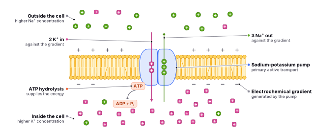
Primary active transport involves the direct use of energy, usually in the form of , to move molecules against their concentration gradient. The sodium-potassium pump is an example of primary active transport, which uses ATP to pump sodium ions out of the cell and potassium ions into the cell. This process is essential for maintaining the resting potential of the cell, as well as for the proper functioning of nerve and muscle cells.
Find it in the figure
Coming soon for figure 4.5
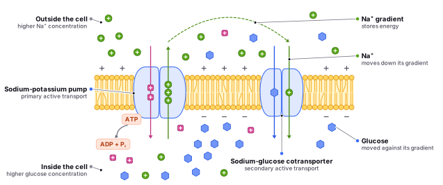
Secondary active transport uses the energy generated from the movement of one molecule down its concentration gradient to transport another molecule against its concentration gradient. An example of secondary active transport is the transport of glucose into the cell in the small intestine. This process involves the movement of sodium ions down their concentration gradient, which generates energy that is then used to transport glucose into the cell.
In short, primary active transport involves the direct use of energy to move molecules against a concentration gradient. In contrast, secondary active transport involves the use of energy generated from the movement of molecules down a concentration gradient to transport other molecules against their concentration gradient.
Primary active transport can be compared to a person using a hand-cranked elevator to move a heavy box to a higher floor. The person must use energy to turn the crank and lift the box against the force of gravity. Similarly, in primary active transport, the cell uses ATP to move molecules against their concentration gradient.
Secondary active transport is like using a pulley system to pull yourself up the hill. The pulley system represents the transport protein, which helps to move you up the hill. However, instead of using energy directly, you are using the energy generated from the movement of another object, such as a heavy bag of rocks, to power the transport. The bag of rocks represents the molecules moving down the concentration gradient (the opposite side of the hill) and generating the energy needed to move you up the hill.
Find it in the figure
Coming soon for figure 4.6
In summary, passive transport occurs without the need for energy and moves molecules down their concentration gradient, while active transport requires energy and moves molecules against their concentration gradient. Primary active transport uses ATP to move molecules, while secondary active transport uses the energy generated from the movement of one molecule to transport another molecule.
The Sodium-Glucose Transporter
The Glucose-Na+ Transporter (also known as the Sodium-Glucose Co-Transporter, SGLT) is a protein that is responsible for the transport of glucose and sodium ions across cell membranes. It is vital in regulating blood sugar levels and electrolyte balance in the body.
There are two main types of SGLT proteins: SGLT1 and SGLT2. SGLT1 is primarily found in the small intestine and is responsible for absorbing glucose and sodium ions from the gut into the bloodstream. SGLT2 is located in the kidneys and is responsible for the reabsorption of glucose and sodium ions from the renal tubules back into the bloodstream.
The SGLT proteins use the energy from the concentration gradient of sodium ions to transport glucose across the cell membrane. When the concentration of sodium ions is higher outside the cell than inside, due to the active pump of sodium, the SGLT protein uses this gradient to transport glucose into the cell. The movement of Glucose is supported by the energy of secondary active transport, as the Sodium ions are actively pumped against their concentration gradient by the ATPase pumps.
In the small intestine, SGLT1 helps to absorb glucose from the diet and maintain normal blood sugar levels. In the kidneys, SGLT2 helps to reabsorb glucose filtered out of the bloodstream by the renal tubules. This helps to prevent the loss of glucose in the urine and maintain normal blood sugar levels.
Defects in the SGLT proteins can lead to problems with blood sugar regulation. For example, mutations in SGLT1 can cause glucose-galactose malabsorption, a condition in which the body cannot absorb glucose and galactose from the diet. On the other hand, inhibiting SGLT2 has been shown to be effective in the treatment of type 2 diabetes, as it helps to lower blood sugar levels by decreasing the reabsorption of glucose in the kidneys.
Quick Final Review
Diffusion is the movement of molecules from an area of higher concentration to an area of lower concentration until the concentration is equal on both sides of the membrane. This process occurs naturally and does not require energy.
Osmosis is the movement of water molecules across a membrane from an area of high water concentration (high water potential) to an area of low water concentration (low water potential). This process also occurs naturally and does not require energy.
Facilitated diffusion is the movement of molecules across a membrane with the help of transport proteins. Like passive transport, facilitated diffusion occurs down a concentration gradient and does not require energy. However, the transport proteins help the molecules cross the membrane more efficiently, allowing them to move against a concentration gradient that would otherwise be too steep for them to cross on their own.
Active transport requires energy and moves molecules against a concentration gradient, while passive transport does not require energy and moves molecules down a concentration gradient. Both processes play important roles in maintaining homeostasis within cells and are essential for the proper functioning of all living organisms.
Bulk Transport
Bulk transport is the process by which cells move large molecules or particles, such as proteins and organelles, into or out of the cell. There are four main types of bulk transport: Endocytosis, Phagocytosis, Pinocytosis, and Exocytosis.
Endocytosis is the process by which cells take in molecules or particles by enclosing them in a vesicle or small membrane-bound sac. There are three types of endocytosis: phagocytosis, pinocytosis, and receptor-mediated endocytosis.
Phagocytosis is the process by which cells engulf and internalize solid particles, such as bacteria or dead cells. This process is often carried out by specialized cells called phagocytes, which are found in the immune system. Phagocytosis occurs when the cell extends a portion of its plasma membrane around the particle, forming a phagosome, which then fuses with a lysosome to form a phagolysosome. Remember that the lysosome contains hydrolytic enzymes that break down the particle into smaller molecules, which can later be absorbed by the cell.
Pinocytosis is the process by which cells take in small amounts of extracellular fluid, along with any dissolved molecules or particles. This process is often referred to as “cell drinking” and occurs when the cell extends small invaginations, or infoldings, in the plasma membrane, forming a pinocytotic vesicle.
Receptor-mediated endocytosis is the process by which cells take in specific molecules or particles by binding them to receptors on the cell surface. The receptors and their associated molecules or particles are then internalized into the cell in a vesicle.
Exocytosis is the process by which cells secrete molecules or particles by expelling them from the cell in a vesicle. This process occurs when a vesicle containing the molecules or particles fuses with the plasma membrane and releases its contents to the outside of the cell. Exocytosis is important for the secretion of hormones, enzymes, and other signaling molecules, as well as for the removal of waste products from the cell.
In summary, bulk transport involves the movement of large molecules or particles into or out of the cell. Endocytosis is the process by which cells take in molecules or particles, while exocytosis is the process by which cells secrete molecules or particles. Phagocytosis, pinocytosis, and receptor-mediated endocytosis are three types of endocytosis that allow cells to internalize specific molecules or particles.